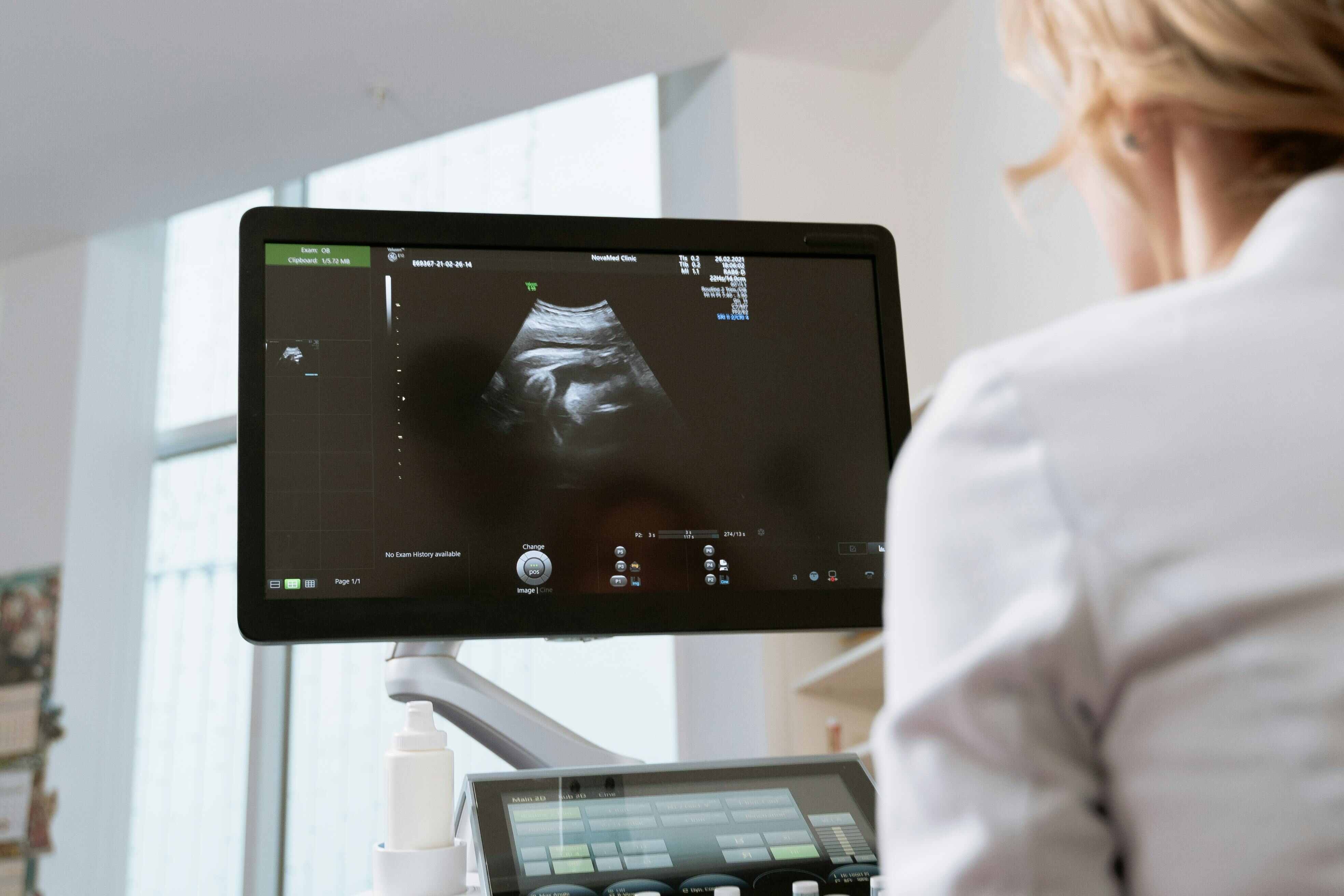
Ultrasound has become an important part of modern cancer diagnosis, particularly when clinicians need a safe, accessible and real-time method of examining soft tissues and internal organs. Unlike X-rays and CT scans, ultrasound uses high-frequency sound waves rather than ionising radiation to produce images of structures inside the body. This makes it particularly useful when repeated imaging may be required or when clinicians need to assess a suspicious lump, mass, cyst, organ abnormality or blood-flow pattern.
The Role of Ultrasound in Cancer Diagnosis extends beyond simply detecting an abnormality. Ultrasound can help distinguish fluid-filled cysts from solid masses, characterise suspicious lesions, assess blood flow, examine nearby structures and guide minimally invasive procedures such as needle biopsies. However, ultrasound is generally one component of a wider diagnostic pathway rather than a standalone test that can definitively establish whether a lesion is cancerous. A definitive diagnosis may require tissue sampling and pathological examination.
Its value lies partly in its flexibility. Ultrasound can be performed relatively quickly, can provide images in real time, does not expose patients to ionising radiation and can be adapted to examine different organs and anatomical regions. Depending on the clinical question, it may be used before other imaging investigations, alongside CT or MRI, or during a biopsy or treatment procedure.
What Is Ultrasound and How Does It Work?
Ultrasound, also known as ultrasonography or sonography, uses a device called a transducer to send high-frequency sound waves into the body. These waves interact with tissues and organs and produce echoes. A computer processes the returning echoes to create images that can be viewed on a monitor in real time.
Different tissues reflect sound waves differently. Fluid-filled structures tend to produce relatively few echoes and can therefore appear darker on an ultrasound image, whereas solid tissues and masses may produce stronger echoes. The resulting differences in appearance help clinicians assess the structure and characteristics of an abnormal area.
The ultrasound probe can usually be moved across the skin after a water-based gel has been applied. For certain examinations, specialised probes may be introduced into a body opening to obtain clearer images of structures that cannot be adequately assessed from the surface. Examples include transvaginal and endorectal ultrasound examinations.
Ultrasound can also incorporate Doppler techniques. Doppler ultrasound evaluates blood movement through vessels and can provide information about the speed and direction of blood flow. Differences in vascularity can sometimes provide additional information when clinicians are assessing a suspicious lesion.
What Is the Role of Ultrasound in Cancer Diagnosis?
The Role of Ultrasound in Cancer Diagnosis varies according to the suspected cancer, the anatomical location of the abnormality and the patient's symptoms and clinical history.
Ultrasound may be used to:
investigate a palpable lump or swelling;
identify an abnormal mass;
determine whether a lesion is predominantly cystic or solid;
examine organs and soft tissues;
assess blood flow within or around a lesion;
evaluate suspicious lymph nodes;
assist with the localisation of a lesion;
guide a needle biopsy;
support certain minimally invasive treatments;
monitor known abnormalities over time;
complement findings from mammography, CT or MRI.
Imaging investigations can be used to identify suspicious areas, determine how extensive a cancer may be and assist with treatment planning. They may also be used to monitor treatment response or investigate possible recurrence.
Ultrasound is therefore best understood as a versatile component of the cancer diagnostic pathway. Its importance is particularly evident when a clinician needs immediate information about a soft-tissue abnormality or needs accurate real-time guidance for a procedure.
Can Ultrasound Detect Cancer?
Ultrasound can identify abnormalities that may represent cancer, but an ultrasound image alone does not always establish whether a lesion is malignant.
For example, ultrasound may reveal a mass and provide information about its size, shape, internal appearance and relationship with surrounding structures. It may also help determine whether the mass is fluid-filled or solid. However, benign and malignant conditions can sometimes have overlapping imaging characteristics. Consequently, further investigation may be required.
This distinction is important because the words “abnormal ultrasound” and “cancer diagnosis” do not mean the same thing.
An abnormal finding may represent:
a benign cyst;
a benign solid tumour;
inflammation;
infection;
scar tissue;
a reactive lymph node;
another non-cancerous condition;
or a malignant tumour.
When imaging raises sufficient concern, clinicians may recommend additional imaging or a biopsy. A biopsy allows cells or tissue to be examined under laboratory conditions and can provide the pathological evidence required for diagnosis.
Ultrasound for Detecting Tumours and Masses
One of the most useful applications of ultrasound is the assessment of a newly discovered lump or mass.
A clinician may request an ultrasound after a physical examination identifies an abnormality. The examination can provide information about whether the structure is fluid-filled or solid and can help determine its precise anatomical location.
This information can influence the next stage of investigation. A clearly benign-appearing cyst may be managed differently from a solid or otherwise suspicious lesion. Where additional assessment is necessary, ultrasound findings can help determine whether further imaging or tissue sampling is appropriate.
Ultrasound can also measure a lesion and document its appearance. When imaging is repeated, clinicians may compare examinations to determine whether an abnormality has changed over time.
Role of Ultrasound in Breast Cancer Diagnosis
Breast ultrasound is an important diagnostic imaging technique, particularly when a breast lump or other abnormality requires further assessment.
Ultrasound can help clinicians examine breast tissue and evaluate suspicious areas. It can also be used to guide a needle during a breast biopsy, allowing the clinician to target the abnormal area accurately.
Breast ultrasound is not necessarily a replacement for mammography. Instead, the two techniques may provide complementary information. The appropriate imaging approach depends on factors such as the patient's age, symptoms, clinical findings and characteristics of the abnormality.
An ultrasound may be especially useful when the clinical question concerns the nature of a palpable lump or when a suspicious area requires targeted assessment.
Ultrasound in Thyroid Cancer Assessment
Ultrasound has an important role in evaluating thyroid nodules.
A thyroid ultrasound can show the structure of the thyroid gland and provide detailed information about nodules within it. Clinicians can assess features such as the nodule's size, composition and appearance and determine whether further investigation may be warranted.
When a thyroid nodule requires tissue assessment, ultrasound can also guide a fine-needle aspiration or other needle-based procedure. The ability to visualise the target in real time allows the needle to be directed toward the intended area.
Ultrasound of the neck may also be used when clinicians are evaluating lymph nodes or investigating possible thyroid malignancy. Ultrasound is one of several imaging methods that may be incorporated into the diagnostic assessment depending on the clinical circumstances.
Ultrasound for Liver and Abdominal Cancer Assessment
Ultrasound is frequently used to examine abdominal organs, including the liver.
When a clinician suspects an abnormality in the liver, ultrasound can help identify focal lesions and other structural changes. Doppler techniques can provide additional information about blood vessels and blood flow.
Ultrasound may also be used when investigating abnormalities involving the gallbladder, pancreas, kidneys and other abdominal structures. However, the usefulness of ultrasound varies between organs and individual patients. Structures containing or located behind air can be difficult to examine because sound waves do not travel effectively through air.
If an ultrasound identifies a suspicious finding, CT or MRI may subsequently be used to provide additional anatomical detail. The choice depends on the organ involved, the suspected condition and the clinical question.
Ultrasound and Lymph Node Assessment
Lymph nodes can become enlarged for many reasons, including infection, inflammation and cancer.
Ultrasound can provide information about the appearance and structure of accessible lymph nodes and can help identify nodes that require further assessment. In selected circumstances, ultrasound can also guide needle sampling of a suspicious lymph node.
This makes ultrasound particularly valuable when the target is superficial and can be visualised clearly with the transducer.
However, the appearance of a lymph node on ultrasound does not automatically establish the presence or absence of cancer. Clinical findings, other imaging and, when indicated, pathological examination must be considered together.
Doppler Ultrasound and Cancer
Doppler ultrasound adds information about blood flow to conventional ultrasound imaging.
Tumours can develop abnormal vascular patterns, and Doppler techniques may help clinicians assess blood flow within or around a suspicious lesion. Doppler ultrasound can therefore contribute additional information during the evaluation of some masses.
Doppler imaging may assess:
direction of blood flow;
relative blood-flow velocity;
vascular patterns;
blood vessels surrounding a lesion;
vascular involvement in selected clinical situations.
Although vascularity can contribute to the characterisation of an abnormality, it should not be interpreted in isolation as proof that a lesion is cancerous.
Ultrasound-Guided Biopsy in Cancer Diagnosis
One of the most clinically significant applications of ultrasound is guiding biopsies.
A biopsy involves obtaining tissue or cells from a suspicious area for pathological examination. When the lesion can be clearly visualised using ultrasound, the transducer can provide real-time guidance as a needle is advanced toward the target.
This can improve targeting and help clinicians sample the intended area rather than relying solely on anatomical landmarks.
Ultrasound-guided biopsy may be used for selected abnormalities involving areas such as the breast, thyroid, lymph nodes and other accessible soft tissues. Radiology guidance is particularly valuable when the abnormality is small or when precise needle placement is required.
Imaging can identify a suspicious lesion, but pathological analysis of a biopsy may be necessary to establish the definitive diagnosis.
Endoscopic and Internal Ultrasound Techniques
Some cancers or suspicious lesions cannot be adequately assessed using a probe placed on the skin. In such situations, specialised internal ultrasound techniques may provide closer access to the target.
For example, an endorectal ultrasound uses a specialised transducer positioned in the rectum. It can help assess how far a rectal cancer has extended through the rectal wall and whether nearby structures or lymph nodes are involved.
Other specialised ultrasound examinations can use probes positioned close to organs or tissues that require detailed assessment. The approach selected depends on the suspected disease and the anatomical region being examined.
Ultrasound for Cancer Staging
Cancer staging determines how extensive a cancer is and where it is located within the body.
Ultrasound can contribute to staging in selected cancers, particularly when clinicians need to evaluate accessible organs, masses, lymph nodes or blood vessels. However, ultrasound is not suitable for every staging question.
CT, MRI, PET and other imaging techniques may provide information that ultrasound cannot obtain, particularly when deeper structures or larger areas of the body need to be assessed. Imaging methods may therefore be combined rather than used independently.
The appropriate investigation depends on the suspected cancer, its location, the information already available and the clinical purpose of the scan.
Ultrasound vs CT Scan for Cancer Diagnosis
Ultrasound and CT provide different types of information.
Ultrasound uses sound waves and does not involve ionising radiation. It is particularly useful for real-time examination of many soft tissues and can be used to guide procedures. CT uses X-rays to create cross-sectional images and can provide a broader and highly detailed anatomical assessment.
Feature | Ultrasound | CT Scan |
|---|---|---|
Energy used | Sound waves | X-rays |
Ionising radiation | No | Yes |
Real-time imaging | Yes | Not in the same way |
Soft-tissue assessment | Useful for many superficial and accessible structures | Broad and detailed anatomical assessment |
Blood-flow assessment | Doppler available | Requires specific techniques/contrast in relevant cases |
Biopsy guidance | Frequently useful for accessible lesions | Also used for image-guided biopsy |
Deep structures | Can be limited | Often more suitable |
Air-filled regions | Limited | Generally more useful |
Cost and accessibility | Often relatively accessible | More complex and resource-intensive |
The choice is not necessarily an “ultrasound versus CT” decision. In many cancer pathways, the examinations answer different clinical questions.
Ultrasound vs MRI for Cancer Diagnosis
MRI uses strong magnetic fields and radiofrequency energy to create highly detailed images of soft tissues. It can provide information that is not available from ultrasound, particularly for deeper or anatomically complex structures.
Ultrasound has several practical advantages, including real-time imaging, portability and the absence of ionising radiation. MRI, however, may be preferred when high-resolution soft-tissue characterisation is required or when the area cannot be adequately evaluated with ultrasound.
For certain tumours, MRI may be particularly valuable for defining the extent of disease and its relationship with surrounding tissues. Consequently, ultrasound can be an initial or complementary investigation rather than the final imaging examination.
Advantages of Ultrasound in Cancer Diagnosis
No Ionising Radiation
A major advantage of ultrasound is that it does not use ionising radiation. This distinguishes it from X-ray-based imaging techniques such as conventional radiography and CT.
Real-Time Imaging
Ultrasound allows clinicians to observe structures dynamically while the transducer is moved. This is particularly useful for guiding needles and evaluating movement or blood flow.
Accessibility
Ultrasound equipment is widely used in hospitals, diagnostic centres and clinical settings. Some systems can also be moved between locations, making ultrasound adaptable to different clinical environments.
Useful for Soft Tissues
Many soft-tissue structures can be examined effectively using ultrasound, particularly when they are close enough to the skin surface for the sound waves to produce useful images.
Procedure Guidance
Ultrasound can guide needles toward selected lesions in real time, making it an important tool for image-guided biopsy and certain minimally invasive interventions.
Repeated Assessment
Because ultrasound does not involve ionising radiation, it can be useful when clinicians need to monitor a known abnormality over time, although the appropriate frequency of follow-up depends on the clinical situation.
Limitations of Ultrasound in Cancer Diagnosis
Despite its advantages, ultrasound has important limitations.
It Cannot Always Confirm Cancer
An ultrasound may identify a suspicious mass but cannot always determine whether it is malignant. Further imaging or biopsy may be necessary.
Bone and Air Can Restrict Imaging
Ultrasound waves do not pass effectively through air or bone. This makes certain anatomical regions difficult to assess and limits the ability to examine structures located behind gas-filled bowel or bone.
Deep Structures May Be Difficult to Visualise
The quality of ultrasound imaging can decrease when the target is deep within the body. Imaging deeper structures can also be more challenging in people with higher body weight.
Operator Skill Matters
The quality of an ultrasound examination can depend on the skill and experience of the person performing the scan. Probe positioning, image optimisation and recognition of relevant anatomy all influence the quality of the examination.
Less Anatomical Detail Than Some Other Imaging
Ultrasound images may not provide the same level of anatomical detail as CT or MRI for particular diagnostic questions.
Can Ultrasound Show the Difference Between a Cyst and a Tumour?
One useful function of ultrasound is helping clinicians distinguish fluid-filled cystic structures from solid masses.
A cyst is generally filled with fluid, whereas a solid lesion contains tissue. The different way these structures interact with sound waves can create recognisably different appearances on ultrasound.
However, this distinction does not mean every solid lesion is cancerous or every cyst is harmless. Some lesions have complex characteristics, and clinical context remains essential.
When an ultrasound finding is indeterminate or suspicious, further imaging or tissue sampling may be recommended.
How Accurate Is Ultrasound for Cancer?
There is no single accuracy percentage that applies to ultrasound for every cancer.
Performance depends on several factors, including:
the type of cancer;
the organ being examined;
the size and location of the lesion;
whether the lesion is superficial or deep;
the patient's anatomy;
the ultrasound equipment;
the experience of the operator;
the imaging technique used;
and whether Doppler or another specialised technique is incorporated.
For this reason, ultrasound should not be described as universally capable of detecting or excluding cancer.
A normal ultrasound can be reassuring in the appropriate clinical context, but it does not necessarily exclude every possible cancer. Conversely, an abnormal ultrasound does not automatically mean that cancer is present.
The interpretation must be integrated with symptoms, physical examination, laboratory findings and other diagnostic investigations.
What Happens After an Abnormal Ultrasound?
An abnormal ultrasound does not necessarily mean that a patient has cancer.
The next step depends on what the examination shows and why the ultrasound was performed. Possible follow-up may include:
Clinical review – the clinician considers symptoms, examination findings and medical history.
Additional ultrasound – a more targeted examination may be appropriate.
CT scan – may provide broader or more detailed anatomical information.
MRI scan – may be selected when detailed soft-tissue characterisation is required.
Biopsy – tissue or cells may be obtained for pathological analysis.
Laboratory investigations – blood or other tests may contribute to the diagnostic assessment.
Follow-up imaging – selected findings may be monitored over time.
Cancer diagnosis is often a process rather than the result of a single examination. Imaging findings are interpreted alongside clinical and pathological evidence.
Does Ultrasound Use Radiation?
No. Conventional diagnostic ultrasound uses high-frequency sound waves rather than ionising radiation. This is one of its principal advantages compared with imaging techniques based on X-rays.
Ultrasound is generally regarded as a very safe diagnostic procedure, with a low risk of adverse effects when used appropriately.
The absence of ionising radiation does not mean every ultrasound examination is identical. Some examinations can involve internal probes or additional techniques, and patients should follow the preparation and procedural instructions provided by their healthcare team.
What to Expect During a Cancer-Related Ultrasound
For a conventional ultrasound, the patient generally lies on an examination table while a trained professional applies gel to the skin over the area being assessed.
The transducer is then moved across the skin to obtain images. Pressure may be applied to improve visualisation, which can occasionally cause mild discomfort, particularly over a tender area.
Patients may sometimes be asked to hold their breath or change position so that particular organs or structures can be visualised more effectively.
Preparation varies according to the type of ultrasound. Some abdominal examinations may require fasting, while certain pelvic examinations may require a full bladder. The healthcare provider will give specific preparation instructions when necessary.
How Long Does an Ultrasound Take?
A typical ultrasound examination may take around 20 to 30 minutes, although the duration varies depending on the area being examined and the complexity of the examination.
A straightforward examination may be relatively quick, while a detailed assessment of a complicated abnormality or an ultrasound-guided procedure may take longer.
The duration should therefore not be interpreted as an indication of whether an examination has found cancer.
The Importance of the Radiology Report
After the ultrasound examination, the images are reviewed and interpreted by an appropriately trained clinician, such as a radiologist, depending on the healthcare setting.
The resulting report communicates relevant imaging findings to the clinician who requested the examination. Radiologists are trained to interpret medical imaging and provide a formal report for the treating team.
A report may describe:
the organs examined;
the size and location of a lesion;
its structural characteristics;
whether it appears cystic or solid;
relevant blood-flow findings;
lymph-node appearances;
comparison with previous imaging;
and recommendations for further assessment where appropriate.
Patients should avoid interpreting an isolated phrase from a report without considering the full clinical context.
The Role of Ultrasound in a Multidisciplinary Cancer Pathway
Cancer diagnosis often involves several healthcare professionals and diagnostic disciplines.
A typical pathway may involve a primary care clinician or specialist identifying a concerning symptom, followed by imaging, specialist review, biopsy and pathological assessment where indicated.
Ultrasound can contribute at several stages:
Initial assessment: identifying or characterising a suspicious abnormality.
Targeted investigation: providing detailed assessment of an accessible structure.
Procedure guidance: directing a biopsy needle toward the lesion.
Staging support: contributing information about selected organs, masses or lymph nodes.
Treatment planning: providing anatomical information that may complement other imaging.
Follow-up: helping monitor selected abnormalities or treatment-related changes.
Imaging may also be repeated during and after treatment to determine whether a tumour has changed or whether there is evidence of recurrence.
Ultrasound and Cancer Treatment Monitoring
Ultrasound is not limited to the initial investigation of a suspicious lesion.
In selected situations, clinicians can use ultrasound to monitor an abnormality over time. Changes in size, structure or blood-flow characteristics may provide useful information when interpreted alongside clinical findings and other investigations.
Imaging is commonly used during cancer care to evaluate treatment response. Depending on the cancer type and treatment, ultrasound may form part of this monitoring strategy, although CT, MRI, PET or other imaging techniques may be more appropriate for particular cancers.
Ultrasound-Guided Cancer Treatment
The real-time nature of ultrasound makes it useful not only for diagnosis but also for selected image-guided procedures.
Ultrasound can guide a needle into a target during procedures such as biopsy. It can also assist with certain tumour-directed interventions, including selected ablation procedures.
The ability to see the target and needle position simultaneously can help clinicians perform procedures accurately while limiting unnecessary tissue disruption.
Is Ultrasound Better Than CT or MRI for Cancer?
There is no universal “best” cancer imaging test.
Ultrasound, CT and MRI have different strengths. Ultrasound is particularly valuable for accessible soft tissues, real-time imaging, Doppler assessment and image-guided procedures. CT provides cross-sectional anatomical imaging using X-rays and is frequently useful when a wider anatomical region must be assessed. MRI provides highly detailed soft-tissue imaging and is particularly useful for certain tumour types and anatomical regions.
The most appropriate test is determined by the suspected cancer, location of the abnormality, clinical question and information already obtained.
In some cases, the most effective diagnostic pathway involves several imaging modalities rather than relying on one.
Frequently Asked Questions About the Role of Ultrasound in Cancer Diagnosis
Can ultrasound diagnose cancer by itself?
Usually, ultrasound identifies and characterises abnormalities rather than providing definitive pathological confirmation. When cancer is suspected, a biopsy and laboratory examination of tissue may be necessary to establish the diagnosis.
Can a normal ultrasound rule out cancer?
Not necessarily. Ultrasound has technical and anatomical limitations, and some cancers may not be adequately visualised. The significance of a normal result depends on the symptoms, clinical examination and type of cancer being investigated.
Can ultrasound detect a tumour?
Yes. Ultrasound can identify many masses and tumours, particularly in accessible soft tissues and organs. However, detecting a mass does not automatically establish that it is malignant.
Does ultrasound show whether a tumour is cancerous?
Ultrasound can reveal characteristics that make a lesion more or less suspicious, but imaging alone cannot always determine whether a tumour is malignant. Further imaging or biopsy may be required.
Is ultrasound safe?
Diagnostic ultrasound does not use ionising radiation and is generally considered a very safe imaging technique.
Can ultrasound detect cancer spread?
Ultrasound can provide useful information about selected masses, organs, lymph nodes and blood vessels, and may contribute to staging in appropriate circumstances. However, CT, MRI, PET and other tests may be needed to evaluate the full extent of disease.
Can ultrasound guide a cancer biopsy?
Yes. When a suspicious lesion can be clearly visualised, ultrasound can provide real-time guidance for needle biopsy.
Does ultrasound use radiation like a CT scan?
No. Ultrasound uses sound waves, whereas CT uses X-rays.
How long does a cancer-related ultrasound take?
Many ultrasound examinations take approximately 20 to 30 minutes, although the duration depends on the body region, examination type and complexity.
Conclusion
The Role of Ultrasound in Cancer Diagnosis is broader than simply locating a tumour. Ultrasound provides real-time imaging, helps characterise masses, differentiates cystic and solid abnormalities, evaluates blood flow and can guide biopsies and selected minimally invasive procedures.
Its lack of ionising radiation, accessibility and ability to visualise many soft tissues make it a valuable diagnostic tool. At the same time, ultrasound has important limitations: it may be less effective for deep structures or areas obscured by air or bone, image quality can depend on operator expertise, and an ultrasound examination cannot always determine whether an abnormality is cancerous.