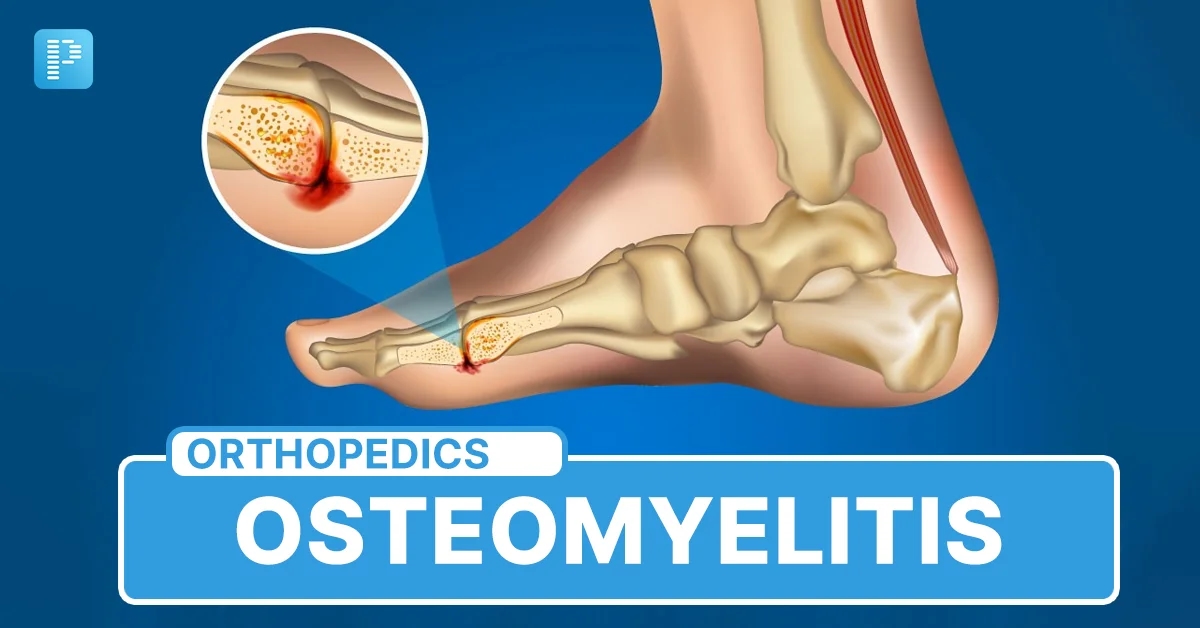
Osteomyelitis is a bone infection that can develop when bacteria or, less commonly, fungi enter bone tissue. Early diagnosis is important because untreated infections may damage bone, affect nearby joints, and cause serious complications. Imaging tests help doctors identify infection and determine how far it has spread.
Although osteomyelitis xray findings can help identify bone damage, magnetic resonance imaging (MRI) is generally more sensitive for detecting early infection. Understanding the differences between these imaging methods can help patients recognise what doctors look for and why additional investigations may be necessary.
This guide explains how bone infection appears on MRI, what early changes look like, how X-rays contribute to diagnosis, and which treatments are used to manage osteomyelitis safely.
Osteomyelitis Xray Findings and the Role of MRI
An X-ray and an MRI provide different types of information about infected bone. An X-ray uses radiation to produce images of bone structure, while MRI uses magnetic fields and radio waves to show bone marrow, soft tissues, and surrounding structures.
Doctors may request both examinations because they answer different diagnostic questions.
How Does Early Osteomyelitis Appear on MRI?
MRI can reveal changes inside bone marrow before substantial bone destruction becomes visible on conventional radiographs. In some cases, abnormalities can be detected within one or two days after infection begins.
Common MRI findings include:
Bone marrow oedema: Fluid accumulation makes infected marrow appear abnormal.
Reduced T1 signal: Normal fatty marrow produces a relatively bright signal on T1-weighted images. Infection replaces some of this normal appearance with darker areas.
Increased T2 and STIR signal: Inflammation and fluid produce brighter areas on fluid-sensitive sequences.
Soft tissue inflammation: Nearby muscles and tissues may become swollen.
Periosteal inflammation: Infection can irritate the membrane covering the outer surface of the bone.
These findings help radiologists identify suspicious areas and assess whether infection extends beyond the bone.
What Does an MRI Report Usually Mention?
An MRI report may describe abnormal marrow signal, surrounding inflammatory changes, cortical involvement, or an abscess. The radiologist considers the location, distribution, and appearance of these abnormalities alongside the patient's symptoms.
Contrast-enhanced MRI may provide additional information about abscesses, infected tissue, and areas of reduced blood supply. However, contrast is not necessary for every examination.
An abnormal MRI does not automatically confirm infection. Bone bruising, fractures, inflammatory conditions, recent surgery, and other disorders can produce similar appearances. Doctors interpret imaging alongside examination findings and laboratory results.
What Does an Early Osteomyelitis Xray Show?
An early X-ray may appear completely normal even when infection is already developing inside the bone. This happens because conventional radiographs primarily demonstrate changes in mineralised bone rather than early inflammation within the marrow.
Nevertheless, osteomyelitis xray examinations remain useful because they provide an initial assessment and help doctors investigate other possible causes of pain.
Early Radiographic Changes
During the initial stages, possible findings include:
Soft tissue swelling around the affected bone.
Blurring of normal tissue boundaries.
Localised swelling near an infected joint.
Occasionally, subtle changes in bone density.
More obvious abnormalities generally develop later. These may include localised osteopenia, cortical destruction, irregular bone margins, and periosteal reaction.
Radiographic abnormalities often take approximately 10–14 days to become visible, although timing varies between patients and depends on the infection. Therefore, a normal examination cannot reliably exclude early osteomyelitis.
Why Do Doctors Still Request X-rays?
Despite their limitations, osteomyelitis xray examinations offer several advantages:
They are widely available in hospitals and clinics.
They are relatively inexpensive compared with MRI.
They help identify fractures, foreign objects, and other bone abnormalities.
They provide a baseline for comparing future changes.
They can reveal advanced bone destruction and certain chronic complications.
Doctors may recommend MRI when symptoms remain suspicious despite an initially normal radiograph.
How Do MRI and X-ray Findings Differ?
MRI and conventional radiography are complementary imaging techniques. Their usefulness depends on the stage of infection and the information the doctor needs.
Feature | X-ray | MRI |
Early infection | Often normal | Can detect early marrow changes |
Bone marrow oedema | Not visible | Usually visible |
Bone destruction | Visible in established disease | Can show associated abnormalities |
Soft tissue infection | Limited detail | Detailed assessment |
Abscess detection | Limited | More effective |
Radiation | Uses ionising radiation | No ionising radiation |
When interpreting an osteomyelitis xray, doctors look for structural abnormalities that suggest infection has progressed. MRI provides a more detailed assessment of marrow involvement and surrounding soft tissues.
Neither examination should be interpreted independently of clinical findings. A radiologist may also recommend CT or nuclear medicine imaging when MRI is unsuitable or when additional information is needed.
What Causes Osteomyelitis to Develop?
Osteomyelitis develops when microorganisms reach bone tissue and trigger an inflammatory response. Bacteria are responsible for most cases, although fungal infections can occur in particular circumstances.
The most common bacterial cause is Staphylococcus aureus. Other organisms may be involved depending on the patient's age, medical history, immune status, and the route through which infection develops.
Three Main Routes of Bone Infection
1. Infection through the bloodstream
Bacteria can travel through the bloodstream from an infection elsewhere in the body and settle in bone. This route is particularly important in children.
2. Direct contamination
An open fracture, penetrating injury, or surgical procedure can introduce microorganisms directly into bone tissue.
3. Spread from nearby infection
An untreated skin ulcer, infected wound, or deep soft tissue infection can extend into neighbouring bone. This is particularly important in people with diabetes and reduced foot sensation.
Who Has a Higher Risk?
Certain conditions increase the likelihood of developing bone infection:
Diabetes, particularly when accompanied by foot ulcers.
Poor circulation and reduced tissue healing.
Recent bone surgery or orthopaedic implants.
Open fractures and serious injuries.
A weakened immune system.
Long-term wounds or recurrent skin infections.
Certain bloodstream infections.
Recognising these risk factors helps healthcare professionals decide whether further investigations are necessary.
What Symptoms Suggest Early Bone Infection?
Symptoms depend on the location, severity, and duration of infection. Acute osteomyelitis may develop quickly, while chronic infection can cause persistent or recurring problems.
Common symptoms include:
Localised bone pain or tenderness.
Swelling around the affected area.
Redness and increased skin temperature.
Fever and chills.
Difficulty walking or moving the affected limb.
Tiredness and a general feeling of illness.
Some patients, particularly older adults and people with diabetes, may experience less obvious symptoms. A foot infection can progress without severe pain when nerve damage reduces sensation.
When Should You Seek Medical Attention?
Seek prompt medical assessment if you experience persistent bone pain accompanied by swelling, warmth, fever, or a nearby infected wound.
Medical warning
High fever, rapidly spreading redness, severe pain, confusion, or signs of sepsis require emergency assessment. Do not wait for an imaging appointment when symptoms suggest a serious infection.
How Is Osteomyelitis Diagnosed?
Diagnosis usually involves several investigations rather than relying on one scan. Doctors consider symptoms, medical history, physical examination, laboratory results, imaging, and microbiological testing.
Blood Tests
Common investigations include:
Complete blood count (CBC): Checks white blood cell levels and other blood components.
C-reactive protein (CRP): Measures inflammation and can help monitor treatment response.
Erythrocyte sedimentation rate (ESR): Provides another indicator of inflammation.
Blood cultures: May identify bacteria circulating in the bloodstream.
Normal inflammatory markers do not completely exclude osteomyelitis. Results must be interpreted in context.
Bone Biopsy and Culture
A bone biopsy involves collecting a small tissue sample for laboratory examination. Microbiological culture can identify the organism responsible and help doctors select an appropriate antibiotic.
Whenever clinically safe, obtaining samples before starting antibiotics can improve the chance of identifying the causative organism. However, treatment must not be delayed in patients who are seriously ill.
An osteomyelitis xray examination may contribute useful information, but a bone culture can provide important microbiological evidence that imaging alone cannot establish.
Can Osteomyelitis Become Chronic?
Yes. Osteomyelitis can become chronic when infection persists, treatment is incomplete, or damaged bone prevents adequate infection control.
Chronic disease may involve areas of dead bone, recurrent inflammation, and persistent drainage through an opening in the skin.
Imaging Features of Chronic Infection
Advanced imaging may demonstrate:
Sequestrum: A fragment of dead bone separated from healthy bone.
Involucrum: New bone formation surrounding an infected area.
Cloaca: An opening through which infected material can drain.
Sinus tract: An abnormal passage connecting an infected area with the skin.
Bone sclerosis: Increased bone density around chronic infection.
An osteomyelitis xray examination can reveal some of these established structural changes, particularly sequestra, sclerosis, and cortical abnormalities.
MRI can provide additional information about active inflammation, soft tissue involvement, and possible abscesses. However, persistent MRI abnormalities after treatment do not always indicate ongoing infection.
Doctors therefore assess clinical improvement, inflammatory markers, and imaging together rather than using one scan as the sole measure of recovery.
What Treatments Are Available for Osteomyelitis?
Treatment depends on the causative organism, infection severity, affected bone, and presence of complications. The main objectives are to eliminate infection, preserve bone function, relieve symptoms, and prevent recurrence.
Antibiotic Treatment
Antibiotics are the primary treatment for most bacterial cases. Doctors select medication according to the suspected organism, culture results, antibiotic sensitivity, and patient-specific factors.
Treatment may involve:
Intravenous antibiotics during initial hospital care.
Oral antibiotics when clinically appropriate.
Several weeks of treatment, depending on the infection.
Follow-up blood tests to monitor response.
Some patients require longer treatment, particularly when infection is complicated or involves chronic bone damage.
Surgical Treatment
Surgery may be necessary when antibiotics alone cannot adequately control infection.
Possible procedures include:
Draining an abscess.
Removing dead or infected bone.
Cleaning contaminated tissue.
Managing infected implants when necessary.
Reconstructing damaged bone or surrounding soft tissue.
An osteomyelitis xray examination may help surgeons understand established structural damage before planning an operation. MRI can help identify the extent of infection and nearby collections that may require drainage.
The treatment plan should be developed by an appropriate medical team, which may include infectious disease specialists, orthopaedic surgeons, radiologists, and wound-care professionals.
Can Natural Approaches Support Bone Infection Recovery?
Natural approaches may support general health during recovery, but they cannot reliably eliminate bacteria from infected bone. Patients should never replace prescribed antibiotics or recommended surgery with home remedies.
Adequate nutrition, hydration, rest, and proper wound care can support the body's normal healing processes.
Some people explore Natural Remedies for Osteomyelitis as complementary wellness measures. However, there is insufficient clinical evidence that herbal preparations or home remedies can cure osteomyelitis. Any supplement should be discussed with a healthcare professional because some ingredients may interact with antibiotics or other medication.
Helpful Recovery Habits
Eat balanced meals containing adequate protein.
Include vegetables, fruits, and whole grains.
Drink sufficient water unless fluid restriction has been advised.
Follow wound-cleaning instructions.
Avoid smoking because it can impair circulation and healing.
Attend follow-up appointments.
Take prescribed medication exactly as directed.
Patients with diabetes should pay particular attention to blood glucose management and daily foot inspections.
What Is the Role of Natural Treatment During Recovery?
Recovery from osteomyelitis requires medical supervision, particularly when infection has affected deeper bone structures. Supportive care can improve general wellbeing, but it should not be confused with antimicrobial treatment.
A Natural Treatment for Bone Infection should never be presented as an alternative to medically prescribed antibiotics. There is no established herbal remedy that can reliably remove a bacterial infection from bone tissue.
Patients interested in complementary approaches should discuss their choices with their doctors before using supplements, essential oils, or traditional preparations. This is especially important for individuals taking multiple medications or recovering from surgery.
Can Nutrition Improve Recovery?
Good nutrition helps maintain muscle strength, supports tissue repair, and provides essential nutrients during recovery.
Important nutritional considerations include:
Protein from eggs, fish, beans, and dairy products.
Calcium from dairy foods and suitable fortified alternatives.
Vitamin D for normal bone health.
Vitamin C from citrus fruits and vegetables.
Zinc from meat, seeds, nuts, and legumes.
These nutrients support normal physiological functions but do not directly cure bone infection.
How Is Recovery Monitored After Treatment?
Recovery varies according to infection severity, the affected bone, treatment response, and underlying medical conditions.
Doctors may monitor patients through regular examinations, blood tests, wound assessments, and additional imaging when clinically indicated.
An osteomyelitis xray follow-up examination may show whether structural abnormalities are stabilising or improving. However, bone remodelling can continue after the infection has been controlled, so radiographic changes may persist.
MRI is not routinely required after every successful treatment course. Doctors generally request repeat imaging when symptoms continue, complications are suspected, or the clinical response is unclear.
Signs That Treatment Is Working
Positive recovery indicators may include:
Reduced bone pain and tenderness.
Decreasing swelling and redness.
Improved movement and daily functioning.
Healing of wounds or drainage sites.
Reduced inflammatory marker levels.
Absence of recurring fever.
Patients should complete their prescribed treatment and report new symptoms promptly.
Can Herbal Care Products Cure Osteomyelitis?
No. Herbal preparations cannot be considered a proven cure for osteomyelitis. Bacterial bone infections require appropriate medical treatment, and delaying effective care can increase the risk of permanent damage.
People researching Herbal Care Products should distinguish between general wellness products and treatments supported by clinical evidence. Natural ingredients may have interesting laboratory properties, but laboratory findings do not establish that a product can reach infected bone, eliminate bacteria, or prevent complications in humans.
Before purchasing supplements, check their ingredients, quality standards, and possible interactions with prescribed medicines. Ask a qualified healthcare professional whether a particular product is suitable for your circumstances.
Frequently Asked Questions
1. Can an X-ray detect osteomyelitis in its early stages?
Usually, early infection is difficult to identify using conventional radiographs. Bone marrow changes develop before substantial mineral loss, so initial images may appear normal. MRI is more sensitive for early detection.
2. What is the earliest sign of osteomyelitis on MRI?
Bone marrow oedema is an important early finding. It generally appears as reduced signal on T1-weighted images and increased signal on T2-weighted or STIR images.
3. How long does osteomyelitis take to appear on an X-ray?
Visible bone abnormalities may take approximately 10–14 days to develop. However, the timing varies depending on the patient, infection location, and disease progression.
4. Can osteomyelitis be present with a normal X-ray?
Yes. Early infection may exist even when radiographs show no obvious abnormalities. Doctors may recommend MRI when symptoms and examination findings continue to suggest bone infection.
5. Is MRI better than X-ray for diagnosing osteomyelitis?
MRI is generally more sensitive for identifying early bone marrow infection and evaluating surrounding soft tissues. X-rays remain useful for initial assessment and identifying established structural changes.
6. Does osteomyelitis always require surgery?
No. Many patients respond to appropriate antibiotics without surgery. However, abscesses, dead bone, infected implants, and certain chronic infections may require surgical intervention.
7. Can osteomyelitis return after treatment?
Yes. Recurrence is possible, especially when infection is difficult to eliminate or underlying problems remain untreated. Follow-up appointments and completing prescribed treatment are important.
8. Is osteomyelitis a serious condition?
It can be serious if treatment is delayed. Infection may destroy bone, spread into surrounding tissues, or enter the bloodstream. Early medical assessment improves the opportunity for effective treatment.
Conclusion
Osteomyelitis requires careful diagnosis because early bone infection may not produce obvious changes on conventional radiographs. MRI provides more detailed information about bone marrow inflammation, soft tissue involvement, and possible abscess formation.
Although an osteomyelitis xray examination remains a valuable diagnostic starting point, a normal result does not exclude infection. Doctors may combine imaging with blood tests, clinical assessments, and bone cultures to establish the diagnosis.
Early treatment, appropriate antibiotics, surgery when necessary, and consistent follow-up are essential for managing this condition. Patients experiencing persistent bone pain, swelling, fever, or infected wounds should seek professional medical assessment rather than relying on home remedies.